Diagnostics
Cardisiography
Cardisiography is an examination that uses artificial intelligence to detect heart disease. It provides a structured check of the heart in a short time.
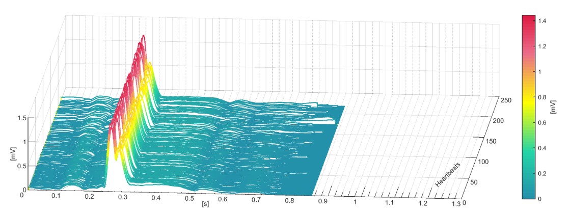
Cardiovascular disease often remains unnoticed for a long time. Symptoms such as chest pressure, shortness of breath or reduced performance may occur only intermittently, and a standard EKG often does not reliably show reduced coronary blood flow. Cardisiography (CSG) is a non-strenuous test that assesses the heart within a few minutes and can help detect abnormalities early.
What is cardisiography?
Cardisiography is a non-invasive examination: only electrodes are placed on the skin; no needles or injections are used. It combines vectorcardiography, which records the heart’s electrical activity in three dimensions, with analysis by artificial intelligence (AI).
A conventional EKG maps the heart in two dimensions. Cardisiography records the heart muscle in three dimensions. This can reveal patterns that a standard EKG may miss.
How is the examination performed?
The examination is straightforward, painless and takes about 10 minutes in total:
- Brief preliminary discussion. We explain the procedure and answer your questions.
- Recording (about 4 minutes). Five electrodes are placed on the chest and body, similar to an EKG. You sit or lie relaxed and remain as still as possible. Exercise on a bicycle or treadmill is not required.
- Analysis. The recorded data are transferred in encrypted form to a secure server and analysed with the AI algorithm. The result is available within a few minutes and is then assessed by a physician.
No special preparation is required. You can resume your usual activities immediately afterwards.
What can it show?
Cardisiography can indicate a number of heart problems, including:
- Reduced blood flow to the heart muscle (ischaemia), as seen in coronary heart disease – including when there are not yet any symptoms.
- Heart rhythm disorders, such as atrial fibrillation or extra beats.
- Signs of structural changes of the heart.
- Indications of an increased risk of a future cardiac event.
Important: cardisiography is a first-assessment method. If it shows abnormalities, we discuss the next steps and arrange further tests if needed (for example ultrasound, MRI or cardiac catheterisation). Conversely, a normal result does not exclude every heart disease with certainty – it is always part of an overall medical assessment.
How accurate is cardisiography?
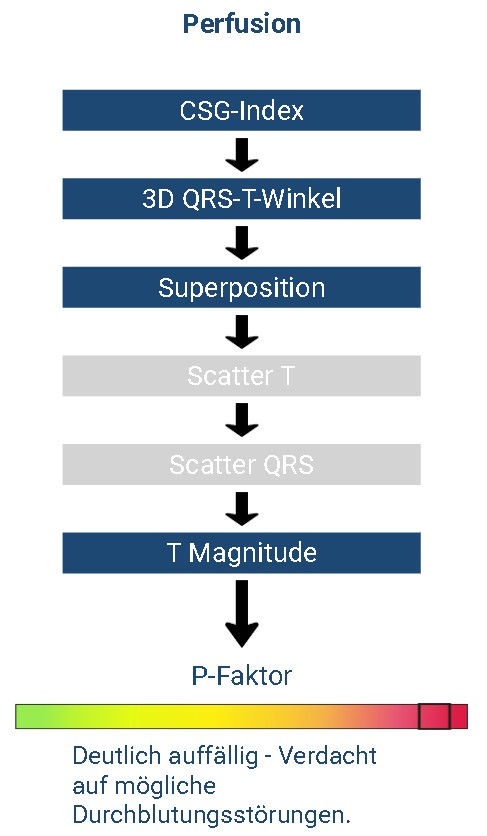
For detecting reduced coronary blood flow, studies and manufacturer data report a sensitivity of more than 90 % for cardisiography. By comparison, the conventional exercise EKG often reaches only about 45–50 %. Cardisiography can therefore identify a large share of affected people without physical exercise.
These figures come from clinical evaluations of the method. The final assessment of each result is always made by Dr. Hierl.
Benefits at a glance
- Gentle and painless – no injections, no radiation, no physical exercise.
- Fast – the whole examination including the result takes about 10 minutes.
- More informative than a resting EKG for reduced coronary blood flow, because the heart is recorded in three dimensions.
- Early detection – abnormalities can become visible before symptoms appear.
- Clarity – you learn promptly whether further cardiac investigation is advisable.
Who may benefit from the examination?
Cardisiography is particularly useful for heart screening – for example with risk factors such as high blood pressure, elevated blood lipids, diabetes, a family history, overweight or smoking – and for unclear symptoms such as chest pressure, shortness of breath or reduced exercise capacity.
Please contact us if you would like advice on whether cardisiography is appropriate for you.