Diagnostics
Echocardiography / TEE
Echocardiography
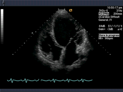
Echocardiography is an ultrasound examination of the heart. It is the main method for assessing heart structure and function and is a standard test when myocardial infarction, heart failure or myocarditis is suspected. Echocardiography is a gentle examination without radiation.
Echocardiography assesses:
- the size of the ventricles and atria
- the function of the heart and the heart valves
- the thickness of the heart muscle
- the pressure in the pulmonary artery
- the pericardium
In transthoracic echocardiography the ultrasound probe is placed on the front of the chest while you lie on an examination couch. The physician directs the probe toward the different parts of the heart to obtain a clear image of the cardiac structures, which are shown on a screen.
When echocardiography is useful
Common reasons are a heart murmur, breathlessness, oedema, reduced exercise capacity, known valve disease, high blood pressure with suspected cardiac strain, or follow-up after infection or chemotherapy.
Preparation and duration
You lie on the examination couch with the upper body uncovered. Gel and the probe are placed on the chest wall. The scan usually takes 15 to 30 minutes, is painless and uses no radiation. Transoesophageal echocardiography (TEE) is a different procedure and is only used for specific questions after separate information.
Related pages
The information on this page does not replace individual medical advice, examination or treatment. Whether any measures are appropriate is decided in a personal consultation. Courses differ from person to person.