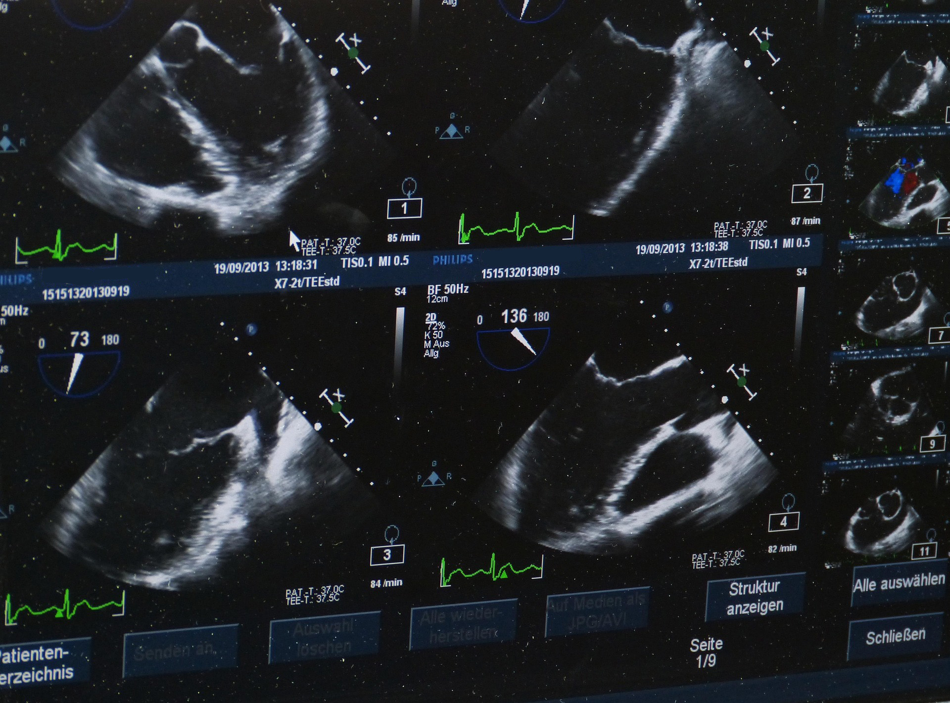
Diagnostics
Abdominal ultrasound
Abdominal ultrasound provides images of the liver, gallbladder, stomach and bowel, lymph nodes, kidneys, spleen, pancreas, bladder, prostate, uterus and ovaries, the abdominal aorta and the blood supply of the organs.
How is the examination performed?
You lie on your back with the abdomen uncovered. After applying gel, the probe is moved over the abdominal wall with light pressure. The examination uses ultrasound waves, is painless and uses no X-rays. You should preferably attend fasting so that the organs, for example the gallbladder, can be assessed more clearly. The examination usually takes about 15 minutes.
What can it show?
Abdominal ultrasound can show many changes of the internal organs:
- Changes of the liver (for example fatty liver) and gallbladder (for example gallstones)
- Kidney stones, cysts and obstruction of the kidneys
- Findings in the spleen, pancreas, bladder and prostate
- enlarged lymph nodes and free fluid in the abdomen
- widening of the abdominal aorta (aortic aneurysm)
How accurate is the examination?
Ultrasound is a fast, safe method that can be repeated as often as needed and is a basic examination in internal medicine. It provides real-time images of the organs and is suitable for early detection and follow-up. For specific questions it can be supplemented by CT or MRI.
Benefits at a glance
- Painless and without radiation.
- Real-time images – many organs in one examination.
- Early detection of, for example, gallstones or an aortic aneurysm.
- Can be repeated as needed – suitable for screening and follow-up.